


Breast cancer remains the most frequently diagnosed malignancy among women in the United States, yet structural obstacles continue to impede early detection and timely medical intervention. Healthcare providers face persistent gaps across the entire care continuum: more than half of women over the age of 40 do not attend recommended annual screenings, while imaging centers confront an impending shortage of tens of thousands of radiologists over the next decade. Even after a suspected tumor is detected, critical molecular and genomic assays that guide therapeutic choices often require weeks to process in specialized external laboratories.
Accelerating Ultrasound Screening Workflows
Addressing the front end of diagnostic care, medical technology startups are re-engineering standard screening workflows to reduce examination times and expand clinical access. Traditional handheld breast ultrasound examinations can take up to 45 minutes and are highly dependent on the manual technique of the individual operator, often preventing direct image comparison across successive annual visits.
To overcome these operator-dependent limitations, iSono Health developed the ATUSA platform, an FDA-cleared wearable system that conducts automated 3D quantitative ultrasound scans. The system captures an entire standardized breast volume in approximately two minutes per breast. Built with specialized GPU acceleration and open-source medical imaging software, ATUSA’s underlying artificial intelligence models were trained on thousands of full-breast scans comprising more than 1.5 million individual ultrasound frames. According to the company, the resulting volumetric scans demonstrate a 28 percent increase in sensitivity over conventional 2D handheld ultrasound.
Because the device secures uniform volumetric views during every session, it provides clinicians with standardized longitudinal data to evaluate tissue changes over time. ATUSA is currently deployed across commercial partner clinics in California, Texas, Georgia, Tennessee, and Washington, D.C. iSono Health is expanding its software to include lesion detection, 3D segmentation, and classification, with long-term plans to integrate multimodal diagnostic intelligence across mammography, MRI, ultrasound, and clinical patient records. A multicenter trial enrolling 3,200 patients is actively evaluating the system, with primary investigation sites located at UC Davis and Vanderbilt University Medical Center.
Triaging Mammograms and Assessing Patient Risk
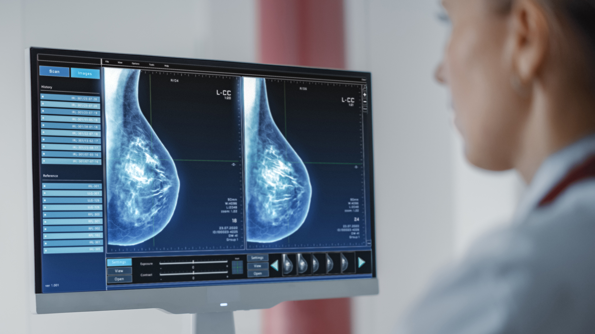
In the mammography reading room, where approximately 40 million examinations are performed nationwide each year, radiologists face a labor-intensive screening process. Clinicians typically locate approximately one malignancy for every 200 mammograms evaluated. Whiterabbit.ai has introduced artificial intelligence software to streamline this high-volume screening workload and assist with individualized risk stratification.
The company’s WRDensity software, which has received FDA clearance, automatically assesses breast tissue density directly from mammograms and has been utilized in clinical care for hundreds of thousands of patients. Whiterabbit.ai has also introduced WRRisk, a clinical decision support system engineered to calculate long-term breast cancer risk. The company is actively researching algorithms capable of detecting malignancies while simultaneously filtering out negative screening mammograms to relieve administrative burdens on radiologists, decrease unnecessary patient recalls, and mitigate downstream medical expenses. The startup trains its diagnostic models on a dedicated GPU cluster situated at Washington University in St. Louis along with supplementary cloud resources, while deploying on-premises computing hardware directly inside participating clinics for real-time inference.
Digitizing Pathology and Forecasting Treatment Efficacy
Following a confirmed diagnosis, oncologists must assess how an individual patient’s tumor will respond to targeted therapies or systemic chemotherapy. Conventional predictive tests frequently depend on separate tissue biopsies sent to outside facilities, generating delays of two to four weeks before treatment can commence.
Ataraxis AI is building software models designed to predict treatment outcomes directly from digital pathology slides and routine clinical variables that are already gathered during standard diagnostic workups. The company has deployed two primary models in active clinical use: one predicts whether preoperative chemotherapy will sufficiently reduce tumor volume prior to surgical intervention, while a second evaluates five-year recurrence probabilities and calculates the therapeutic benefit of postoperative chemotherapy. Validated across clinical trials and more than 10 medical institutions, these models are executed across on-premises servers, offsite data centers, and cloud infrastructure using the PyTorch framework accelerated by specialized parallel computing architectures.
Multimodal Precision Modeling for Surgical Planning
Complementing digital pathology advances, SimBioSys applies precision medicine technology to create patient-specific 3D anatomical visualizations. By processing MRI scans, tumor pathology, and clinical records, the company builds spatial models of breast tumors, surrounding vascular networks, and adjacent soft tissues to assist surgeons during operative planning.
SimBioSys also utilizes volumetric data to calculate the probability of cancer recurrence. The company leverages medical imaging AI frameworks such as MONAI alongside high-performance computing libraries to analyze complex imaging datasets in the cloud, allowing clinical teams to access multiparametric modeling data without waiting days or weeks for external laboratory results. While certain tools across these early-stage ventures remain investigational and await commercial FDA clearance, their deployment underscores a broader transition toward high-throughput computational tools across oncology.